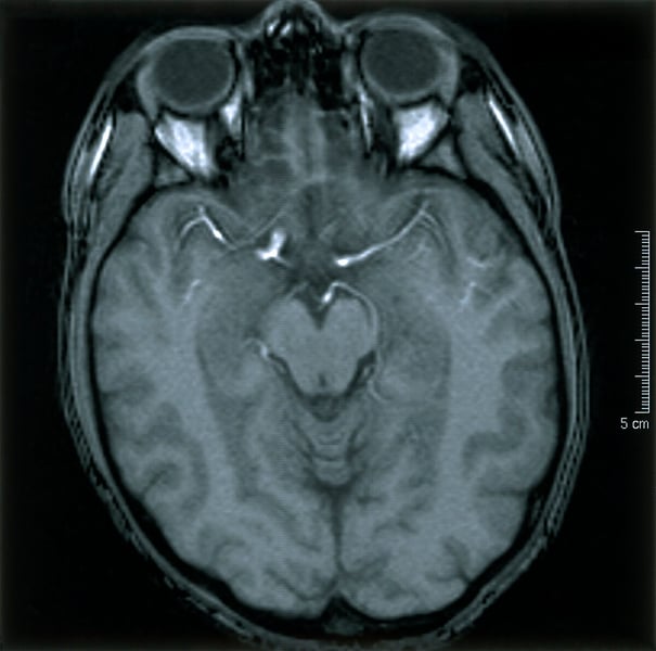

OSLO — For decades, the clearest answer a doctor could offer someone worried about Alzheimer’s disease was a negative amyloid PET scan: no plaques visible, no cause for clinical alarm. A study published in Nature Neuroscience now complicates that reassurance. In people who later developed Alzheimer’s-related amyloid accumulation, the brain was already changing in ways detectable by a standard MRI, not by months but by at least seven years before any elevated protein appeared on the gold-standard scan.
The research, led by James Michael Roe at the Center for Lifespan Changes in Brain and Cognition at the University of Oslo, combined 4,570 longitudinal MRI scans with 1,684 amyloid PET scans drawn from three separate cohorts of cognitively healthy adults. The team found that structural MRI, measuring the thickness of the brain’s outer layer, the cortex, can detect a pre-disease signature more than seven years earlier than the imaging technology medicine has treated as the benchmark for identifying Alzheimer’s risk. The researchers published their analysis, in the study, in Nature Neuroscience in August 2026.
The finding was counterintuitive. People who went on to develop elevated amyloid did not show early cortical thinning, as the Alzheimer’s literature would predict in a brain already under biological stress. They showed a thicker cortex and less cortical thinning over time compared with those who remained amyloid-negative. A brain on the path toward Alzheimer’s disease was operating measurably differently from a healthy brain years before it began to visibly deteriorate, and that difference expressed itself, paradoxically, as excess tissue in the cortex rather than its loss.
Roe, who conducted the research at Oslo’s LCBC and now serves as International Scientific Lead at Cercare Medical, described the implication directly: structural changes in the brain occur many years before high levels of plaque are seen on PET scans, which is the brain scan currently used to identify the earliest signs of Alzheimer’s disease.
The finding’s credibility rests partly on the scale of the dataset. The team drew on scans from three independent, cognitively healthy cohorts, tracking the same participants longitudinally with MRI data collected at least seven years before each person’s first amyloid-positive PET result. Co-authors included William J. Jagust and Susan M. Landau of the University of California Berkeley and Theresa M. Harrison, whose prior work on amyloid conversion informed the study design. The combined dataset, encompassing 4,570 MRI acquisitions alongside 1,684 amyloid PET scans, has no prior equivalent in studies examining pre-amyloid brain structure.
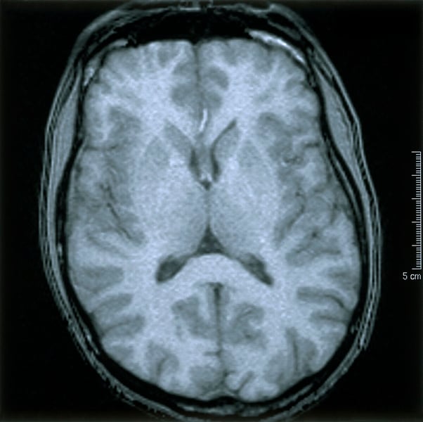
What drives the paradoxical cortical thickening remains unexplained by the paper. Researchers have proposed that it may reflect a compensatory reorganization in neurons under early metabolic stress, before the degeneration that produces visible thinning begins. The study identifies the structural signature; it does not explain the biology that produces it. That gap is the next research question.
The clinical implications depend on a question the paper cannot resolve: whether catching the cortical signature seven years earlier would translate into better patient outcomes. No treatment has been proven effective at this pre-amyloid stage of Alzheimer’s disease. The study proposes no protocol changes. What it does suggest is that amyloid PET, for all its diagnostic value, is drawing the starting line for medical intervention later than the brain’s actual biology would warrant.
That finding arrives at a difficult moment for Alzheimer’s medicine. The case for rethinking when the disease begins has been building from multiple directions. A landmark clinical review found that amyloid-clearing drugs delivered benefits most investigators described as trivial, developed within a framework that treated amyloid accumulation as Alzheimer’s earliest measurable event. The Oslo paper suggests that framework may be missing the first chapter of the disease’s natural history.
Parallel research is assembling a richer picture of how Alzheimer’s operates at the cellular level. Scientists at the University of Utah identified, in July, the Arc protein tau mechanism by which tau, the misfolded tangles most directly linked to cognitive decline, is packaged into vesicles and carried between neurons, providing the first precise account of how the disease travels through brain tissue once established. A separate line of inquiry has been building evidence on early sugar restriction and dementia risk, suggesting that Alzheimer’s risk may be shaped decades before any clinical test is ordered.
Across these inquiries, the gap between detection and intervention persists. The World Health Organization noted, in its fact sheet on dementia, that the approximately 55 million people currently living with dementia worldwide will nearly triple to 153 million by 2050 as the global population ages. Most receive a diagnosis years after the disease has already done substantial structural work.
The Oslo study does not propose a clinical pathway. What it establishes is that a brain destined for Alzheimer’s disease is already distinguishable by MRI, available in virtually every hospital, a full seven years before the imaging benchmark medicine trusts most would have flagged any problem. Whether that seven-year window is long enough to matter, for a drug not yet developed or an intervention point not yet established, is the question this finding leaves open and urgent.
Related Posts