
Protein fibers in semen can help RNA enter sperm cells and reach mouse embryos after fertilization, researchers have found, revealing an unexpected transport role for a molecular structure more commonly associated with brain disease.
The study, published in Molecular Cell on October 26, identifies a route by which genetic messages outside sperm can travel into an embryo. The findings do not establish a new fertility treatment or show that these fibers affect development during natural conception.
William Mallen and Anthony Fitzpatrick at Columbia University’s Zuckerman Institute and their colleagues investigated how amyloid fibers interact with RNA in seminal fluid. Their experiments combined molecular analysis, detailed imaging and laboratory fertilization using mice.
Not the same proteins involved in Alzheimer’s
Amyloid describes the structure proteins adopt when they assemble into fibers. Although the term is familiar from research on Alzheimer’s and other neurodegenerative diseases, an amyloid structure does not automatically mean a protein is causing disease.
The fibers examined here were built from a fragment of prostatic acid phosphatase, or PAP, an enzyme produced by the prostate. These are different proteins from those that accumulate in Alzheimer’s disease, ScienceAlert reported. Seminal amyloid fibers can form outside cells in healthy men and can be taken apart again.
That distinction matters because the research concerns a possible normal biological function, not evidence that Alzheimer’s-associated proteins are being transmitted through sperm.
RNA helps assemble its own carrier
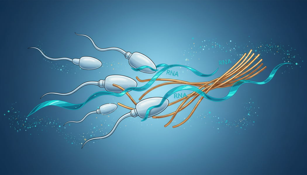
The team studied positively charged PAP fragments and negatively charged RNA. When mixed in the laboratory, they bound together, formed small droplets and then assembled into fibers containing RNA.
Without RNA, the PAP fragments remained in solution under the conditions tested. Adding RNA brought them together and accelerated fiber formation, suggesting the cargo itself helped build the structure that carried it.
The researchers also found small RNA molecules in an amyloid-containing fraction of human seminal fluid. Many identifiable sequences were fragments of transfer RNA, which ordinarily participates in protein production. That observation connected the laboratory work to material present in semen, without proving how that material affects an embryo.
Imaging confirms fibers enter sperm
To distinguish fibers inside sperm from fibers simply attached to the surface, the researchers tagged laboratory-made fibers with tiny gold particles. They used bull sperm for detailed imaging because those cells are relatively large.
Cryo-electron tomography, which produces three-dimensional images of frozen samples, revealed labeled fibers within sperm. Other microscopy and live-cell imaging supported that finding, while separate experiments demonstrated uptake by human and mouse sperm.
According to Columbia’s account of the study, the work addressed a longstanding question: how RNA in seminal fluid reaches sperm. RNA can carry instructions for proteins or help regulate gene activity, making its route into reproductive cells relevant to early development.
Mouse embryos use the delivered instructions
In laboratory fertilization experiments, mouse sperm exposed to fibers made from human PAP fragments produced two-cell embryos containing those fragments. The team also used RNA labeled with heavier carbon and nitrogen atoms to track delivery. More labeled RNA reached embryos when fibers were present.
A further experiment tested whether the cargo remained functional. Researchers used RNA encoding a green fluorescent protein. Embryos produced from sperm given RNA with the fibers contained at least ten times as much of the marker protein as embryos produced from sperm given the same RNA without fibers.
The result measures production of a laboratory marker. It does not mean that fertility, embryo health or pregnancy success increased tenfold.
Natural effects and safety remain unanswered
Separate laboratory tests found that clusterin, a protein also detected in embryos, accelerated RNA release from the fibers. Its role in releasing that cargo inside embryos remains unconfirmed.
The embryo experiments used mice, laboratory-made fibers and added RNA. No human embryos or natural conceptions were tested. The researchers have not established whether naturally occurring fibers carrying their usual RNA cargo change embryo development.
What happens to the fibers after they enter an embryo is also unknown. The study therefore offers a mechanism to investigate and a possible foundation for future RNA-delivery research, not a demonstrated reproductive therapy.